

Quick answer: A dandruff flake is a cluster of shed skin cells. Magnification can reveal its overlapping layers, but the appearance depends on the instrument and sample preparation. A close-up picture alone cannot reliably diagnose the cause of a flaky scalp.1,2
A magnifying glass, a microscope slide, and a scanning electron micrograph show different levels of detail. Understanding that difference makes the images below much more useful.
The appearance of magnified dandruff
At low magnification, a loose flake looks like an irregular piece of scale. More detailed imaging can show flattened skin cells called corneocytes overlapping in layers. The visible flake is a collection of shed cells, not a living organism.1,3
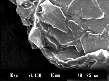
The 10 μm scale bar helps show how small these overlapping layers of shed skin cells are. Both scientific images in this article are real electron micrographs; the cover is an AI-generated editorial illustration of an optical microscope.
The contents of a dandruff flake
The scalp's outermost skin layer, the stratum corneum, continually renews itself. Usually, shedding is too subtle to notice. In dandruff, altered skin renewal and cohesion produce visible clusters. Research has also found changes in the scalp's skin barrier and in cell maturation.1,4
A sample may contain skin oils and material from nearby hairs or products. Neither its color nor its thickness can, by itself, tell you whether the problem is dandruff or dry scalp. The skin's appearance, symptoms, and history matter too.2
Different instruments reveal different details
Scalp magnification
A magnifying lens or dermatoscope shows scale, hair shafts, and the scalp surface. It helps a clinician examine the pattern of a rash but does not provide the same view as a prepared microscope slide.2
Light microscopy
A prepared skin or hair sample can be examined for specific features. A clinician may order fungal testing when another condition, such as scalp ringworm, is suspected.5
Scanning electron microscopy
SEM shows fine surface structure in a specially prepared sample, as in the photograph above. It is a research technique, not a routine home test for dandruff.3,4
Biopsy microscopy
A biopsy examines a small tissue sample rather than just a loose flake. A dermatologist occasionally uses it when the diagnosis is unclear or more than one condition may be present.6
Malassezia and the limits of a flake image
Malassezia is a yeast normally found on skin. Its interaction with scalp oils and a person's skin response plays a role in dandruff and seborrheic dermatitis. Finding it in skin scrapings does not, on its own, diagnose either condition.2
Visible white flakes are not masses of yeast that you can identify with the naked eye. Researchers use appropriately prepared samples and specialist methods to investigate microorganisms and skin structure. A consumer scalp-camera image cannot tell you which yeast species is present.
Research comparisons and diagnostic limits
Research can document differences, but an online picture comparison is not a diagnostic test. The study behind the six-panel image below examined 20 people across several scalp disorders, including only two with dandruff. Its findings should not be turned into a rule for diagnosing everyone with flakes.3
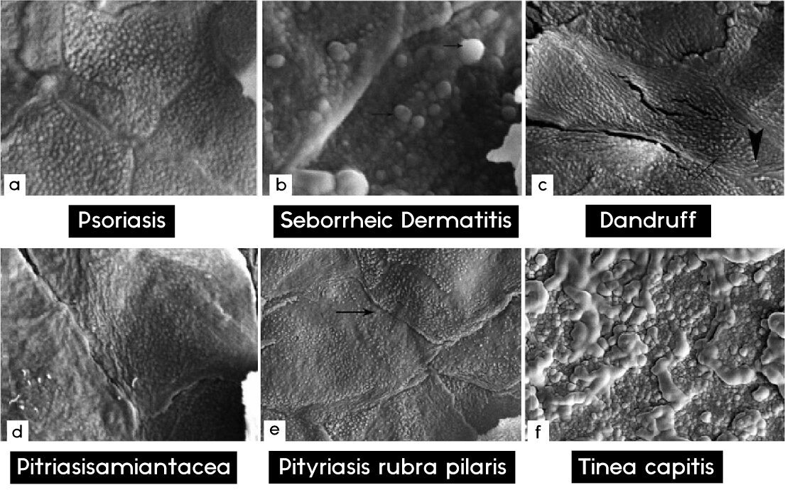
Most dandruff and seborrheic dermatitis assessments begin with the history and scalp examination. Testing is chosen to answer a particular question, not simply because a flake is visible.6
Scalp changes that need assessment
- Patchy hair loss or broken hairs: These can occur with scalp ringworm, which needs medical treatment.5
- Painful swelling, pus, or a very inflamed scalp: Arrange a prompt assessment, particularly in a child.5
- Persistent flaking despite appropriate care: A dermatologist can assess whether dandruff, psoriasis, contact dermatitis, or another condition is responsible.6
Caring for dandruff without a microscope
A dandruff shampoo can help control recurring flakes. Our dandruff guide covers symptoms and care in more detail. Choose an appropriate active ingredient and follow the product label or your clinician's directions.7
Happy Cappy Medicated Shampoo & Body Wash contains pyrithione zinc 0.95% to help relieve flaking, scaling, and itching associated with dandruff and seborrheic dermatitis. Stop use and ask a doctor if the condition worsens or does not improve with regular use as directed.8