


A dark patch on the cheek can have several explanations. It may follow a healed acne spot, appear gradually after years of sun exposure, or develop as part of melasma. Those changes can look similar at first, but their history matters: a mark left exactly where a pimple healed tells a different story from patches that appear across both cheeks during pregnancy.
The distinction is not between two unrelated conditions. Hyperpigmentation describes the color change; melasma names one particular condition that causes it. Understanding that relationship makes it easier to judge which advice applies to your skin.
Hyperpigmentation describes several kinds of darkening
Melanin is the pigment that contributes to normal skin color. Hyperpigmentation develops when more pigment is produced or accumulates in an area, making it look darker than the surrounding skin. Pigment may be present in the epidermis, the outer skin layer, or deeper in the skin. Brown and gray-brown are common appearances; deeper pigment can look slate gray or blue-gray. It does not have to reach a deeper layer before a mark becomes visible.2,3
Pink or red marks after acne are not automatically hyperpigmentation. They may reflect lingering redness, called post-inflammatory erythema, while a flat brown mark reflects a pigment change. An indented or raised acne scar is a change in skin texture. Redness, pigment changes and scars can occur together, but they are not interchangeable terms.
Dark marks after inflammation
Post-inflammatory hyperpigmentation, often shortened to PIH, appears after inflammation or injury. Acne, an eczema flare, an insect bite or irritation from a skin-care product can leave a darker area after the original problem settles. The mark often follows the location of that earlier inflammation. PIH can affect any skin tone, although it is often more noticeable and persistent in darker skin.2,3
For example, a child whose itchy eczema patch has healed may still have a darker area in the same place. That color change does not, by itself, mean the eczema remains active or that the skin has scarred. Our article on eczema marks, color changes and scars explains that distinction in more detail.

Sun-related spots and other causes
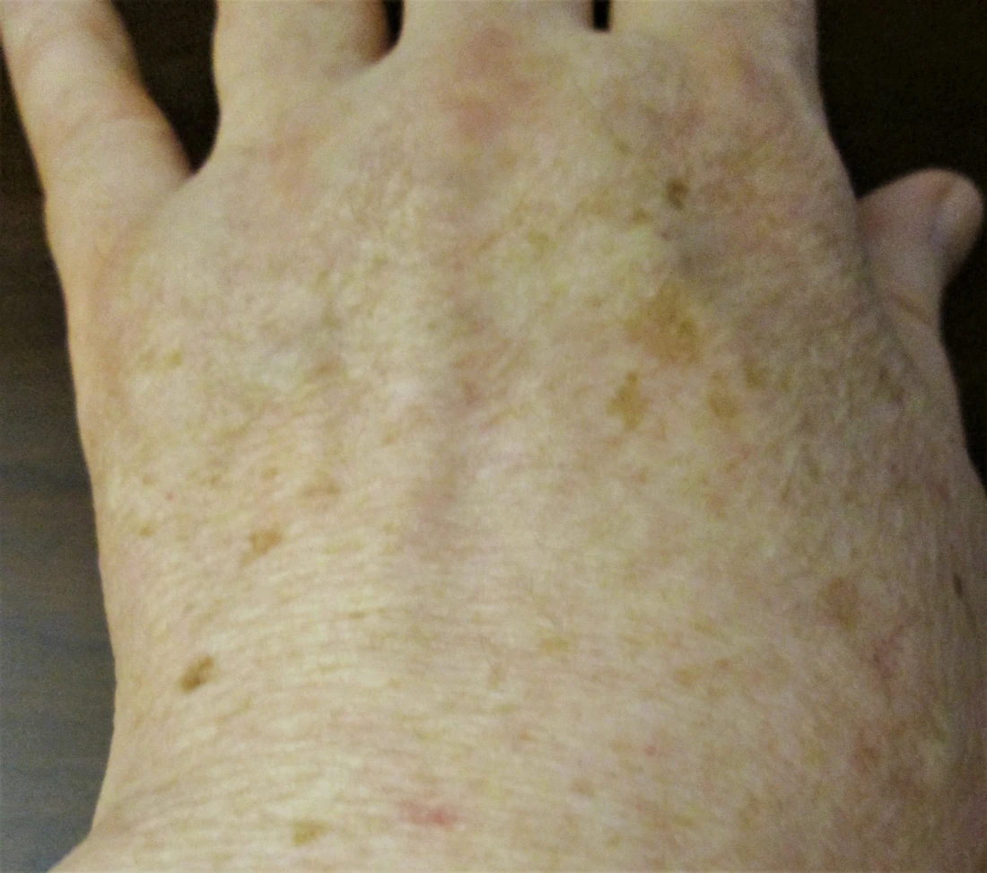
Solar lentigines, often called sunspots or age spots, are usually distinct, flat spots on areas with accumulated sun exposure, such as the face and backs of the hands. Freckles are another pigment pattern and can become more noticeable in sunlight. Medications and some medical conditions can also alter skin color, so a new pattern should not automatically be labeled sun damage or melasma.4

Melasma has a characteristic pattern and set of triggers
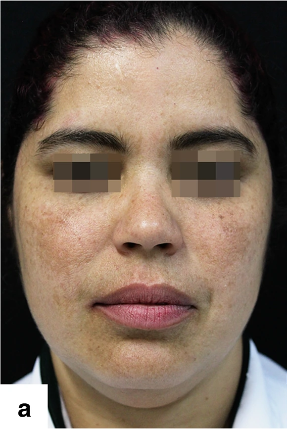
Melasma commonly produces irregular brown or gray-brown patches across both sides of the face. The cheeks, forehead and skin above the upper lip are frequent locations; the nose and chin may also be involved. The patches are generally flat, without a change in the skin's surface. Melasma itself usually does not itch, hurt or scale.1
Clinical photographs of melasma
Two clinical examples show melasma across the cheeks and forehead. These photographs illustrate appearance; a diagnosis also depends on the history and examination.

Melasma occurs more often in women and in people with medium to darker skin tones, but men and people with lighter skin can develop it too. Pregnancy and hormone-containing contraceptives can contribute, alongside a person's susceptibility and exposure to light. It is not exclusively a pregnancy condition, and the presence of melasma does not establish a hormonal imbalance that needs testing or treatment.5
Ultraviolet light can make melasma more noticeable. Visible light from sunlight can also worsen pigmentation, particularly in darker skin tones. This helps explain why patches may deepen after outdoor exposure and why a treatment plan usually includes ongoing light protection. Irritation from aggressive skin care can create additional dark marks, complicating the picture.6,7
Pigment patterns provide clues, not a diagnosis by photograph
The most helpful starting point is the combination of appearance and timing. Consider where the change began, whether a rash or injury came first, and whether the pattern has changed with sunlight, pregnancy or a medication. More than one cause can be present on the same face.
| Feature | Melasma | Post-inflammatory hyperpigmentation | Sunspots |
|---|---|---|---|
| Usual pattern | Irregular patches, often on both sides of the face | Marks where inflammation or injury occurred | Individual, usually well-defined flat spots |
| Helpful history | Light exposure, pregnancy or hormonal medication may contribute | A preceding acne spot, rash, bite or irritation | Accumulated sun exposure over time |
| Course | Often persistent or recurrent | May gradually fade after the cause is controlled | Often remain unless treated |
| Care priority | Consistent light protection and a tailored pigment-treatment plan | Control the original inflammation and avoid further irritation | Confirm the diagnosis before treating an individual spot |
These are typical patterns, not a checklist that confirms a diagnosis. A dermatologist can assess the distribution, examine individual lesions and, when useful, use a dermatoscope or other tools. An unusual lesion may need additional testing before any attempt to lighten it.8
Compare the clinical photographs of melasma, post-inflammatory pigmentation and sunspots above. All three are forms of hyperpigmentation.
Care starts with the cause and consistent light protection
Sun protection supports every pigment-treatment plan
Use a broad-spectrum, water-resistant sunscreen with SPF 30 or higher on exposed skin, alongside shade and a broad-brimmed hat. Reapply sunscreen at least every two hours outdoors and after swimming or sweating, following its label. For melasma, a tinted sunscreen containing iron oxides adds protection against visible light. A product being mineral-based does not, by itself, guarantee that added visible-light protection.6,7
The goal is to limit new stimulation of pigment while existing discoloration improves. Scrubbing cannot wash melanin away. If an exfoliant or other product causes burning, stinging or a rash, continuing it may add inflammation and leave more discoloration.
Treatment follows the specific pigment problem
With PIH, controlling acne, eczema or another source of inflammation helps prevent new marks. Existing marks often take months to fade, and deeper pigment may last longer. Melasma can also take months to improve and may return after it has faded. Improvement after pregnancy is possible, but it is not guaranteed.2,8
A dermatologist may recommend topical treatments such as azelaic acid, prescription hydroquinone, or a prescribed combination of hydroquinone, tretinoin and a corticosteroid for melasma. These medicines have different purposes and precautions. A corticosteroid is not a stand-alone dark-spot remedy, and stronger or more frequent application is not automatically better. In the United States, hydroquinone skin-lightening products are not approved for over-the-counter sale.8,9
Chemical peels and laser or light procedures may help selected patients, but they can also worsen pigmentation. The decision depends on the diagnosis, skin tone and prior treatment response. A procedure suitable for an individual sunspot is not automatically suitable for widespread melasma.
Pregnancy changes medication choices
During pregnancy, prioritize gentle skin care and sun protection. Retinoids and hydroquinone should be avoided; discuss any pigment-lightening medication with your obstetrician or dermatologist before using it. If a prescribed contraceptive or another medicine might be contributing to melasma, discuss alternatives with the prescriber rather than stopping it yourself.10
Moisturizer supports comfort rather than removing pigment
A gentle cleanser and a moisturizer can help dry or easily irritated skin tolerate a sensible routine. They serve a supporting role alongside treatment of the underlying cause. Happy Cappy Moisturizing Cream can be considered for dry, sensitive skin; its role here is moisturization. It is not a treatment for melasma or dark spots, and it does not replace sunscreen.
An ingredient's presence in a moisturizer is not proof that the finished product will lighten pigmentation. For this comparison, the useful product question is whether your everyday care keeps skin comfortable without adding irritation.
New or changing pigmentation deserves the right assessment
Arrange an examination for a new, changing or unexplained dark patch, especially an individual spot that grows, bleeds, develops an irregular border or looks different from your other spots. Melasma does not turn into skin cancer, but other lesions can be mistaken for a harmless pigment change.4
Itching, pain or scaling suggests an additional or different skin problem rather than uncomplicated melasma. Widespread new darkening, medication-related changes or discoloration accompanied by other symptoms also warrants medical assessment. Once the cause is clear, you can choose care that fits it and set realistic expectations for improvement.
References
- American Academy of Dermatology. Melasma: Signs and symptoms.
- American Academy of Dermatology. How to fade dark spots in darker skin tones.
- Davis EC, Callender VD. Postinflammatory hyperpigmentation: a review of the epidemiology, clinical features, and treatment options in skin of color. Journal of Clinical and Aesthetic Dermatology. 2010;3(7):20–31.
- American Academy of Dermatology. What can get rid of age spots?
- American Academy of Dermatology. Melasma: Causes.
- American Academy of Dermatology. Melasma: Self-care.
- Castanedo-Cazares JP, et al. Near-visible light and UV photoprotection in the treatment of melasma: a double-blind randomized trial. Photodermatology, Photoimmunology & Photomedicine. 2014;30(1):35–42.
- American Academy of Dermatology. Melasma: Diagnosis and treatment.
- US Food and Drug Administration. FDA works to protect consumers from potentially harmful OTC skin lightening products.
- American Academy of Dermatology. Dermatologist-approved pregnancy skin care.
Hero artwork: AI-generated editorial illustration with the official Happy Cappy logo; not a clinical photograph.